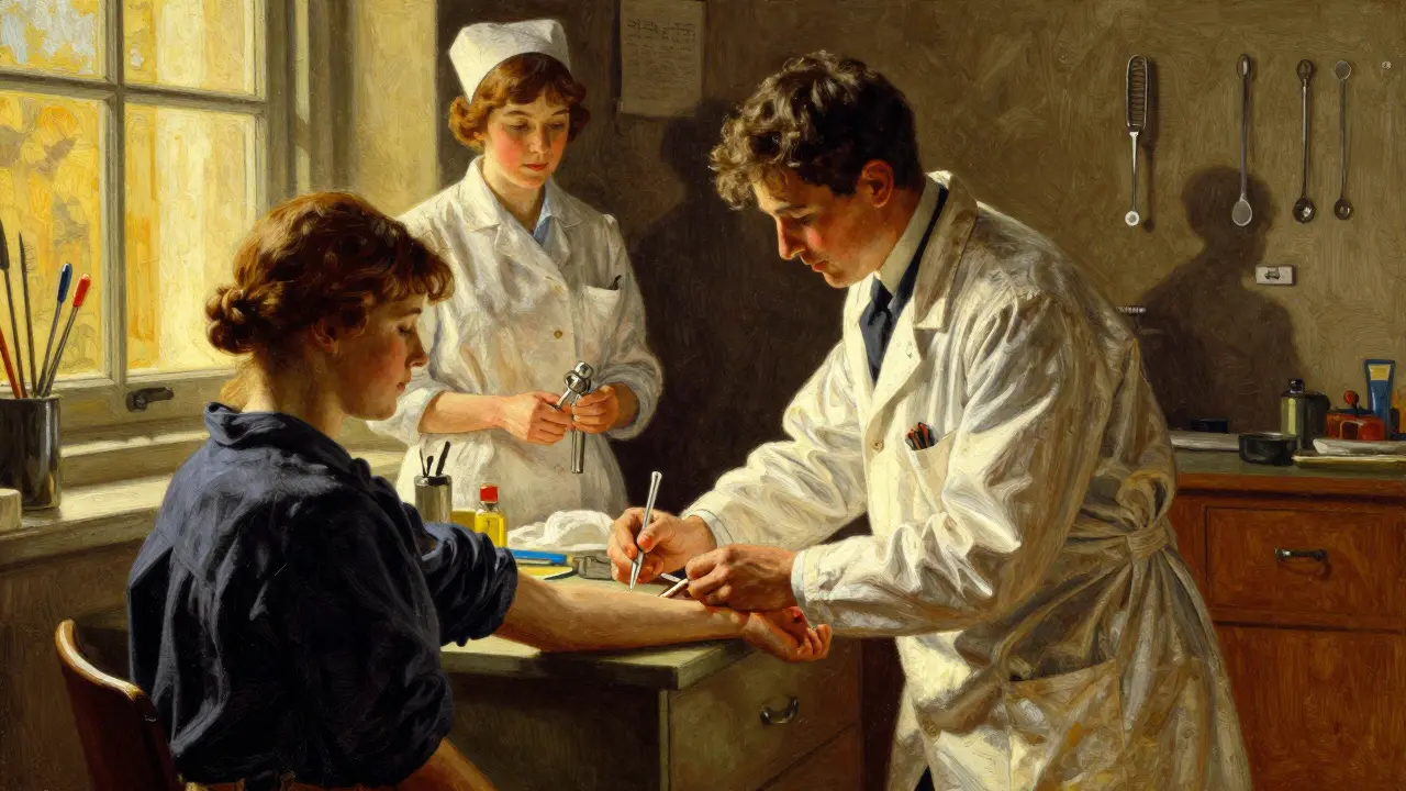
Imagine being able to track cancer progression without another needle sticking into your lung or liver. For decades, doctors relied on tissue biopsies-cutting out a piece of the tumor to study it under a microscope. While effective, that method has limits. It is painful, risky, and often misses the changing nature of the disease. That is where liquid biopsy steps in. By analyzing tiny fragments of genetic material floating in the blood, oncologists can now see inside the body through a simple blood draw.
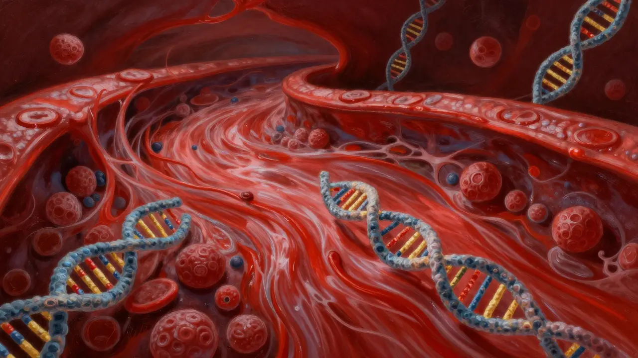
This technology centers on circulating tumor DNA (ctDNA) is tumor DNA released into the bloodstream when cancer cells die.. As a biomarker, it provides real-time data on what is happening inside the tumor. In recent years, major institutions have integrated this approach into standard care pathways, shifting how we handle everything from initial diagnosis to post-surgery surveillance. Understanding this shift is crucial for anyone navigating modern oncology.
Understanding Circulating Tumor DNA
When cancer cells break down, they release their genetic material into the circulatory system. We call these fragments circulating tumor DNA. Unlike normal cell turnover, which produces background noise, tumor-derived DNA carries specific mutations unique to the cancer itself. Detecting these mutations allows doctors to identify the genetic 'fingerprint' of the tumor without ever touching the lesion directly.
The concept isn't entirely new. Research on circulating components began focusing on circulating tumor cells (CTCs) before expanding to include ctDNA and extracellular vesicles. However, ctDNA has emerged as the most versatile component. Studies published in 2024 indicate that ctDNA assays serve critical roles in screening high-risk patients and diagnosing tumor progression dynamically. The key difference lies in abundance and stability; unlike whole cells, DNA fragments persist long enough to be captured in standard plasma samples.
To detect these signals, laboratories use several sophisticated methods:
- Digital Droplet PCR (ddPCR): Highly sensitive, capable of detecting one mutant molecule among 10,000 wild-type molecules.
- Next-Generation Sequencing (NGS): Allows for scanning thousands of genes simultaneously for rare mutations.
- Methylation Profiling: Analyzes chemical modifications on DNA that vary by tissue origin, improving source identification.
- Nanopore Sequencing: Emerging tech that reads long DNA fragments for better structural variant detection.
Liquid Biopsy Versus Traditional Tissue Biopsy
Why switch from the traditional method? Tissue biopsies remain the gold standard for initial pathology, but they face a significant challenge called heterogeneity. A single snapshot might miss mutations present in other parts of the tumor. Research suggests single-site biopsies can miss up to 30% of molecular alterations due to this spatial variation. A blood test, however, captures DNA shedding from all active tumor sites, providing a more comprehensive view of the disease biology.
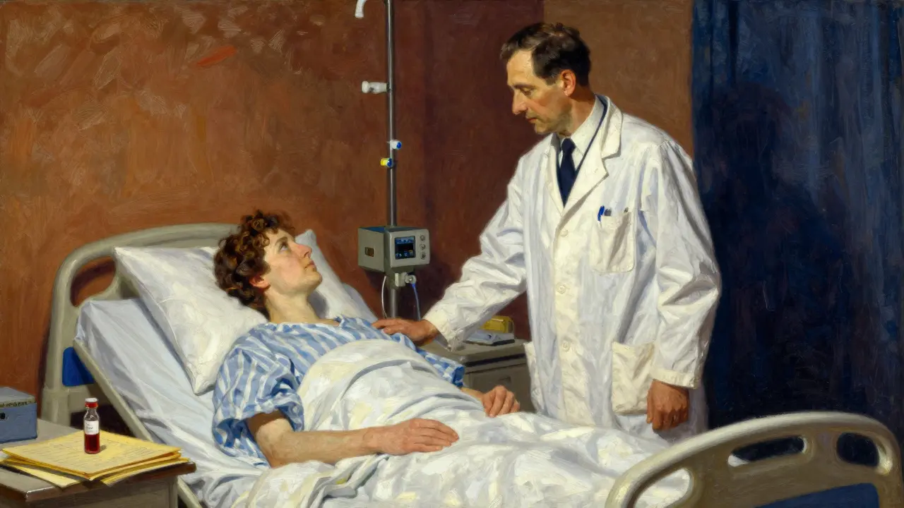
Consider the practical implications for a patient undergoing chemotherapy. With tissue biopsies, you cannot easily repeat the procedure every few weeks to check if the drug is working. With liquid biopsy, you can draw blood monthly. This capability helps distinguish true progression from pseudoprogression-a phenomenon seen in 5-10% of immunotherapy cases where tumors appear larger due to immune infiltration rather than actual growth.
| Feature | Tissue Biopsy | Liquid Biopsy |
|---|---|---|
| Invasiveness | Surgical or needle-based procedures | Blood draw only |
| Heterogeneity Capture | Limited to sampled site | Systemic view of all tumor sites |
| Frequency | Occasional (risky to repeat) | Frequent (every 4-8 weeks possible) |
| Complication Risk | 1-5% depending on tumor location | Minimal |
| Sensitivity in Stage I | High (if accessible) | Varying (50-70%) |
Clinical Applications in Modern Oncology
The utility of ctDNA analysis goes far beyond just seeing cancer. It transforms how we manage the entire treatment lifecycle. One of the most promising areas is detecting minimal residual disease (MRD). After surgery, the naked eye might suggest no cancer remains, yet microscopic traces often linger. ctDNA testing offers 85-90% sensitivity in spotting these remaining traces, predicting recurrence 6-11 months earlier than standard imaging scans.
Another vital application involves identifying resistance mutations. When a targeted therapy stops working, the tumor often mutates again. Dr. Jie-Qun Ma notes that ctDNA analysis can flag these changes up to 3-6 months before radiographic evidence appears. This window gives clinicians time to switch therapies before the patient feels significantly worse. For example, in non-small cell lung cancer, checking for EGFR mutations via blood has become standard when tissue samples are insufficient, occurring in over 90% of such cases successfully.
We also see value in personalized therapy selection. Some patients have 'cold' tumors that don't shed much DNA, while others like colorectal cancers shed heavily. Understanding shedding rates helps interpret results accurately. Furthermore, distinguishing between clonal hematopoiesis (age-related blood mutations affecting roughly 10-15% of people over 65) and true tumor variants is a critical step in reporting to avoid false positives.
Current Adoption and Regulatory Landscape
As of early 2026, the regulatory framework supporting liquid biopsy has matured significantly. The U.S. Food and Drug Administration granted over a dozen approvals between 2020 and 2023, including companion diagnostics like Guardant360 CDx. These approvals provide insurance coverage pathways that were previously nonexistent. Major guidelines, including those from ASCO (American Society of Clinical Oncology), updated recommendations in 2023 to support liquid biopsy for initial biomarker testing in advanced lung cancer.
Market dynamics reflect this trust. The industry is projected to grow rapidly, moving towards widespread integration. Academic centers report that 60-70% of oncology divisions now offer testing regularly. Community practices are catching up, though adoption varies based on cost and reimbursement policies. Approximately 35-40% of phase I clinical trials now incorporate ctDNA as a primary biomarker, signaling a shift in how new drugs are validated.
Limitations and Practical Challenges
Despite the excitement, liquid biopsy is not a perfect replacement for tissue sampling in every scenario. Sensitivity remains a hurdle for early-stage cancers. Detection rates hover around 50-70% for stage I disease compared to near 90% for stage IV. This limitation exists because small tumors simply do not release enough DNA into the bloodstream to pass current detection thresholds.
Standardization is another persistent issue. Different labs use different sequencing panels and algorithms, leading to inter-laboratory variability affecting up to 25% of test results in multicenter studies. Blood collection tubes, processing timeframes, and storage conditions all alter the integrity of the sample. Without harmonized pre-analytical protocols, results can be inconsistent across different providers.
Cost remains a barrier for universal access. While prices are dropping as technology scales, high-throughput sequencing still represents a significant expense for healthcare systems. Additionally, interpreting variants of unknown significance occurs in about 15-20% of reports. This ambiguity requires expert tumor board review, adding a layer of complexity to clinical decision-making that busy physicians must navigate carefully.
Future Directions and Integration
Looking ahead, the integration of artificial intelligence is reshaping fragment analysis. Algorithms are learning to recognize subtle fragmentation patterns and nucleosome positioning that human analysts miss. MD Anderson researchers suggest AI could boost diagnostic accuracy by 15-20% by combining methylation data with fragmentomics. Long-term viability assessments position liquid biopsy to become the standard of care for cancer monitoring within five to seven years.
We are also moving toward multi-analyte approaches. Future tests will likely combine ctDNA with protein markers and extracellular vesicles to create a composite risk score. This strategy aims to push early detection sensitivity above 95%. Ultimately, the goal is a surveillance system that replaces routine CT scans with blood tests, reducing radiation exposure and unnecessary imaging costs by 20-25%.
Is liquid biopsy covered by health insurance?
Coverage varies significantly by region and insurer. In the US, many plans cover FDA-approved companion diagnostics like Guardant360. In the UK and Europe, coverage depends on national health service guidelines, which are increasingly adopting these tests for specific cancers like lung and breast.
Can liquid biopsy replace surgery for diagnosis?
Not currently. While it is excellent for monitoring, a tissue sample is usually still required for initial histological confirmation and staging. Liquid biopsy complements rather than fully replaces tissue biopsy at this stage.
How often should I get tested?
Frequency depends on your situation. During active treatment, testing might occur every 4-8 weeks. During surveillance after surgery, every 3-6 months is typical. Your oncologist will determine the optimal schedule based on tumor type and risk factors.
What happens if my result is 'undetectable'?
An undetectable result generally indicates no measurable circulating tumor DNA. It suggests low tumor burden or effective treatment response. However, 'negative' does not guarantee zero cancer cells, as some tumors shed very little DNA.
Does this work for all types of cancer?
It works best for solid tumors like lung, breast, and colorectal cancers. It is less effective for brain tumors and certain blood cancers where ctDNA shedding rates are naturally lower or confined to specific fluids.


Jordan Marx
looking at the fragmentomics and nucleosome positioning really changes things in analysis
Rohan Kumar
big pharma wants your blood lol 🧐 not sure i trust these stats completely though
Debra Brigman
the essence of healing shifts with every drop of fluid flowing through us
Sabrina Herciu
It is fascinating how the technology is evolving rapidly in our field. We really need to understand the limitations involved though. Standardization remains a massive hurdle for everyone currently. Labs often run different algorithms which causes variance issues. Blood collection tubes matter significantly for results accuracy. Processing timeframes can degrade sample integrity very quickly. You cannot ignore the cost barriers facing patients today. Insurance coverage pathways are still developing globally now. Many people do not know what variants mean without professional help. Interpreting unknown significance is tricky for doctors daily. False positives are a constant worry for affected families always. Clonal hematopoiesis confuses the data quite frequently nowadays. Age-related mutations affect about ten percent of senior citizens. Experts must review every single ambiguous report with care. This complexity requires dedicated attention from medical boards constantly.
This is why we must stay vigilant.
tyler lamarre
how quaint you think a simple blood test solves all the deep issues
Philip Wynkoop
just glad we are seeing progress here :)
kendra 0712
Wow! This is such amazing news!!! We really need hope!!!
Monique Louise Hill
You should really listen to your body and share responsibly 🌸
Devon Riley
Sending good vibes to anyone going through this treatment 😊💪